Ask a CT technologist what determines image quality, and you'll get an answer about the scanner. Ask an ultrasound specialist, and you'll get an answer about the person holding the probe. That gap — between a machine-driven exam and an operator-driven one — is exactly where AI ultrasound is trying to earn its place.
Why Two Operators See Two Different Exams
Ultrasound is a real-time, interactive imaging method. Image quality isn't set by a fixed gantry and preset protocols the way it is in CT or MRI; it's produced by three decisions the operator makes in the moment: where to place the probe, how much pressure to apply, and what the returning echo actually means.
Where the Subjectivity Comes From
The same thyroid nodule can produce two very different images depending on who's scanning — a senior specialist or a first-year resident. This isn't a malfunction; it's the defining property of the modality. The technical term is operator dependence: the acquisition itself carries human judgment, unlike CT and MRI, where the machine reconstructs from a standardized protocol.
What Low Consistency Costs
When different readers can't agree on the same finding, we call it low inter-observer agreement. It doesn't stay abstract — it turns into two concrete costs:
- Unnecessary biopsies. When a reader isn't sure, the safe move is to biopsy, subjecting the patient to a fine-needle aspiration (FNA) they may not have needed.
- Missed findings. When a reader isn't confident, they may also let a suspicious nodule pass, delaying the next step.
⚠️ Watch Out: Low consistency is a systemic property of ultrasound, not a defect in one machine or one clinician. Anyone who claims AI can "eliminate" operator variability is oversimplifying the problem.
How AI CAD Standardizes Nodule Assessment — TI-RADS, BI-RADS, and S-Detect
To see what AI ultrasound is for, start with the thing it targets: risk stratification of thyroid and breast nodules.
Why Risk-Stratification Systems Need Help
TI-RADS (Thyroid Imaging Reporting and Data System) and BI-RADS (its breast counterpart) break a nodule into scorable features — shape, margin, taller-than-wide ratio, echogenicity, microcalcification — and map them to a malignancy risk level. The logic is clean; the execution is the hard part, because those features have historically been eyeballed one at a time, slowly and subjectively.
CAD (computer-aided detection) exists to hand that manual scoring to an algorithm.
S-Detect: Automated Feature Extraction and a Malignancy Hint
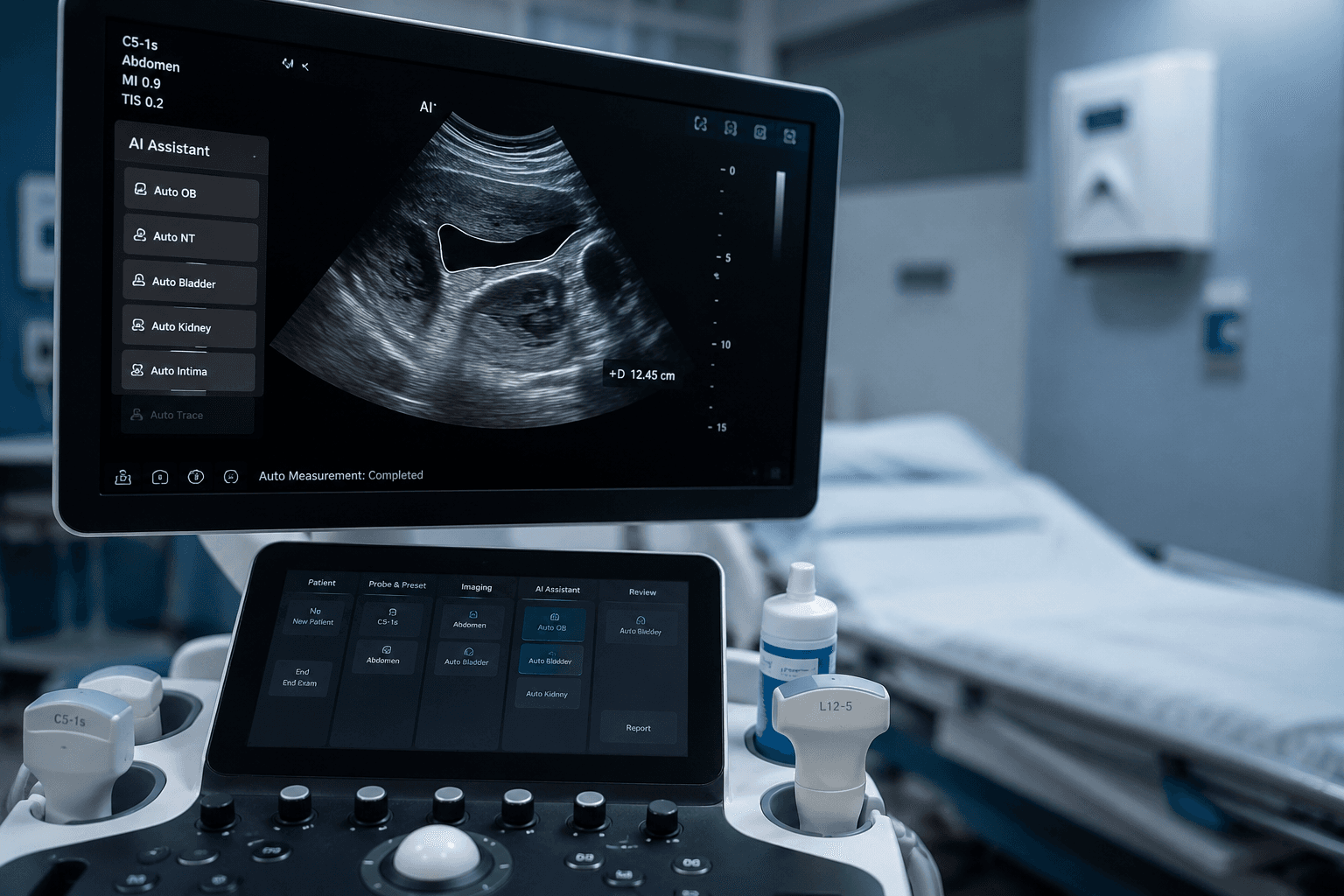
Samsung Medison's S-Detect is the representative example. Integrated into the underlying architecture of its high-end systems, it lets the operator lock onto a nodule, then automatically extracts the lesion boundary and outputs a structured analysis — shape, orientation, margin clarity, echo pattern, microcalcification — along with a probability of malignancy and a follow-up suggestion.
One caveat belongs right here: the 93% nodule classification accuracy S-Detect reports is Samsung's official figure, not an independent third-party verification.1 Keep those two things separate when you evaluate it.
The Effect on Consistency and Unnecessary FNA
Several clinical comparison studies — most of them from vendor training material and review articles, so treat them as low-grade evidence — report that this kind of CAD shortens exam time and narrows the consistency gap between junior and senior readers, which in turn cuts some unnecessary biopsies.2 Read these as "vendor/review claims," not settled, independently verified conclusions.
💡 Expert Insight: The real value of CAD here isn't "more accurate than the expert." It's "closer to the expert's floor for the beginner." It raises the floor, not the ceiling. — If you're pairing this with a hardware decision, our ultrasound transducer buying guide walks through what to check on the probe itself.
Reshaping OB/GYN Ultrasound Workflow — Sonio's Cloud Reports and Visual Checklists
Nodule assessment is a single-point judgment. Obstetric ultrasound is a systemic workflow — and AI intervenes here in a different way.
The Fetal-Anomaly-Scan Problem
A mid-pregnancy anomaly scan means acquiring dozens of standard planes and measuring a long list of fetal structures. Under clinical pressure, with a poorly positioned fetus or a tired sonographer, it's easy to miss a required plane or mis-measure a structure.
Real-Time Checklists, Best-Frame Extraction, Auto-Filled Reports
Platforms like Sonio reframe the process as something closer to "navigation with live safety prompts":3
- A visual checklist monitors the probe's field of view in real time and verifies whether the current image meets the diagnostic-grade plane;
- the system extracts the best frame from the live video stream;
- it auto-detects and fills in core report fields like fetal anatomy parameters and placental position.
To be precise: this is the vendor's functional description. What AI optimizes here is whether the workflow misses or mislabels anything; the clinical diagnosis still belongs to the physician.
Regulatory Milestones and the Push Down-Market — See-Mode, POCUS, Teleradiology
The next chapter for AI ultrasound is moving it out of specialist suites and into broader, less-resourced settings.
The 2024 Regulatory Milestone
In 2024, See-Mode Technologies received FDA 510(k) clearance for its AI thyroid ultrasound analysis software — a milestone for AI in screening superficial organs like the thyroid.4 510(k) is the FDA pathway that establishes a new device is "substantially equivalent" to one already on the market.
Edge AI + Remote Reading, Down to the County and the ER
For county emergency rooms, community clinics, and remote facilities, the bottleneck was never the ultrasound machine — it's the shortage of specialist sonographers. POCUS (point-of-care ultrasound) combined with on-device AI is changing that: a junior physician, a general practitioner, even a nurse, can follow the AI's on-screen guidance to complete a standardized sweep; the AI flags lesions and produces preliminary measurements, and difficult cases are uploaded to a referral center for expert review.4
A related proof point in portable hardware is MIT's $300 portable 3D breast ultrasound, which applies the same down-market logic to breast screening.
The question a buyer should actually ask isn't "does this machine have AI," but "can the AI help my team scan to standard when I don't have a specialist on hand." If you're shortlisting ultrasound systems, you can search by brand, model, and use case on MedTrade and send an inquiry directly to suppliers — browse ultrasound equipment.
The Real Limits of AI Ultrasound — It Doesn't Replace You, and It Isn't Always More Accurate
Once the value is on the table, the boundaries have to go on the table too — otherwise this reads as a vendor pitch.
False Positives and Over-Reliance
AI suggestions can produce false positives, and a clinician who leans too hard on the prompt may end up ordering invasive follow-up that wasn't needed. AI is an assist, not an arbiter.
Cross-Population Generalization
A model trained on one population can degrade on a different population, a different machine, or different scanning habits. This is a general AI-imaging problem, not one unique to ultrasound — our reality check on AI medical imaging digs into the generalization failures in more depth.
The Evidence-Grade Caveat
Most of the numbers in this article — accuracy rates, consistency gains, FNA reductions — come from vendor figures or review articles, which is a lower evidence grade than independent validation. In a purchasing decision, treat these as "vendor claims" until you see independent data.
The bottom line: AI ultrasound's value is real, but it's a standardization tool, not a diagnostic substitute. It nudges ultrasound's defining weakness — operator dependence — toward reproducibility, raising the floor in under-resourced settings without ever replacing the physician's ceiling.
If you're equipping an ultrasound department, a screening center, or a county facility, the practical move is to put "AI capability" on your checklist next to probe model, use case, and supplier credentials — then compare options and send inquiries directly to suppliers on MedTrade: search ultrasound equipment.