Mammography has been the gold standard for breast cancer screening for over 40 years. It is also, by any honest assessment, a deeply imperfect tool. It uses ionizing radiation on healthy women. It performs poorly in the 40–50% of women with dense breast tissue — the very population at elevated risk. And it requires a $100,000–$300,000 machine installed in a dedicated room with a licensed technologist to operate it. These three constraints — radiation exposure, dense-tissue blindness, and centralized equipment monopolies — mean that breast screening remains, for hundreds of millions of women worldwide, either inaccessible, inaccurate, or both.
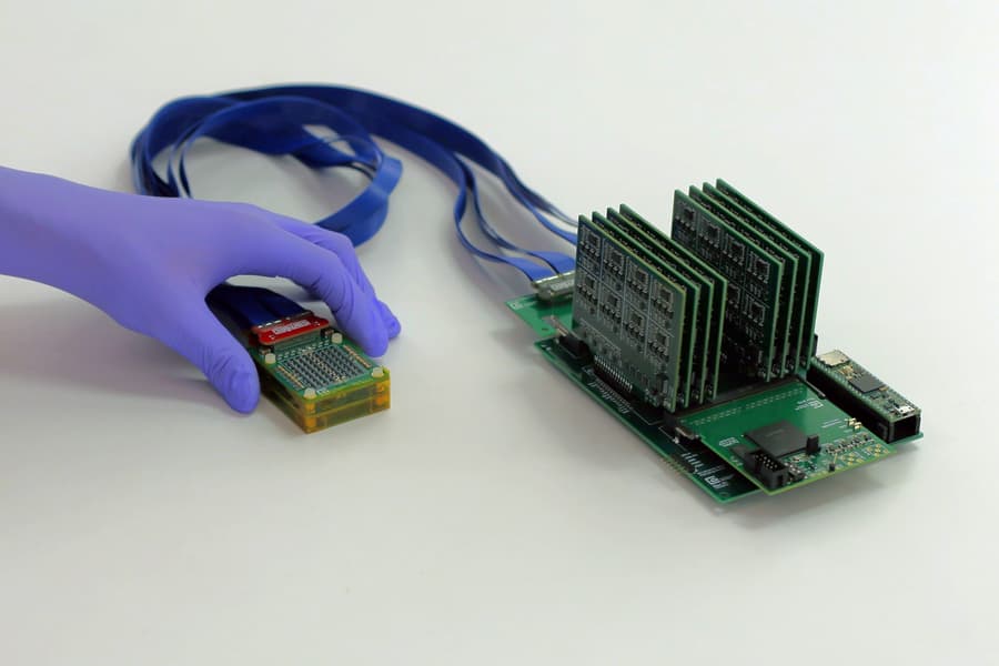
On July 1, 2026, a team at the MIT Media Lab led by Canan Dagdeviren published a paper in Nature Communications describing a device that takes aim at all three problems simultaneously. It is called 3D PURE — a portable, real-time, three-dimensional breast ultrasound system. The hardware is the size of a deck of cards plus a smartphone-sized processing board. The bill of materials cost is approximately $300. And the system is designed to be operated by the patient herself, with no clinical training required.
This article examines the engineering behind 3D PURE, the clinical logic of self-operated breast ultrasound, and what this device — still in pre-trial research, not yet FDA-cleared — signals about the next decade of breast screening.1
The Three Unsolved Problems in Breast Screening
To understand why 3D PURE matters, it helps to be clear about what's broken.
Radiation: the cumulative burden. A screening mammogram delivers a low dose of ionizing radiation — roughly 0.4 mSv, equivalent to about seven weeks of natural background exposure. For a single exam, this is negligible. But screening is not a single exam. Women who begin annual screening at 40 will undergo 30–40 mammograms over their lifetime. The cumulative radiation exposure is non-trivial, particularly for women with BRCA mutations or other genetic risk factors where radiation sensitivity is heightened. Ultrasound has no ionizing radiation. It has always been the safer modality — but it has never been the accessible one.
Dense breasts: the 50% blind spot. Approximately 40–50% of women have heterogeneously or extremely dense breast tissue. In these women, mammographic sensitivity drops from roughly 85% to as low as 47–62%. Dense fibroglandular tissue appears white on a mammogram. So do tumors. Reading a mammogram of a dense breast is, in the words of one radiologist, "like looking for a polar bear in a snowstorm." Ultrasound, by contrast, differentiates tissue by acoustic impedance rather than X-ray attenuation — it sees through density. The challenge has always been operating it.
Equipment monopoly: screening stays inside hospital walls. A modern 3D tomosynthesis mammography system costs $250,000–$400,000. A high-end cart-based ultrasound system adds another $100,000–$200,000. Both require dedicated physical space, a licensed technologist, and a radiologist for interpretation. For women in rural areas, low-resource countries, or even busy urban professionals who skip annual exams, the friction of scheduling, traveling, and waiting is enough to delay or forgo screening altogether. The result is interval cancers — tumors that appear between scheduled screenings and account for a significant fraction of breast cancer mortality. Dagdeviren's team has cited a personal motivation: the lab director's mentor's relative died of an interval breast cancer — a tumor that emerged between annual screenings and was caught too late.1
Inside 3D PURE: Three Layers of Engineering Innovation
The device consists of two components: a square-box probe roughly the size of a deck of playing cards, and a signal-processing board slightly larger than a smartphone. It connects to a laptop via USB. That is the entire system. The engineering breakthroughs are in the acoustic array design, the transducer materials, and the beamforming algorithm.
Corner-Gap Offset Box Array: 128 elements, 1,024-channel equivalent. Conventional ultrasound arrays use a linear or phased arrangement of piezoelectric elements. To achieve high-resolution 3D volumetric imaging, you typically need a 2D matrix array with a large element count — 1,024 or more channels — which drives up cost, complexity, and power consumption. The 3D PURE team took a different approach. Their Corner-Gap Offset Box Array uses only 128 physical elements arranged in a specific geometry where the transmitter and receiver elements are separated — transmitters at the corners, receivers along the edges — creating an acoustic stereo-imaging effect. Through this architecture, 128 physical elements achieve imaging performance equivalent to a 1,024-channel 2D matrix array. The system requires only 2–3 scan positions to cover a full unilateral breast volume.1
Flowable Backing Layer: crosstalk down 4.5 dB, resolution up 200 μm. In any ultrasound transducer, the backing layer behind the piezoelectric elements absorbs rearward-propagating acoustic energy to prevent it from reflecting back into the tissue and creating artifact. The MIT team developed a self-formulated flowable backing material that conforms precisely to the array geometry during fabrication. The result: inter-element crosstalk reduced by 4.5 dB. Axial resolution improved by 200 μm. Lateral resolution improved by 70 μm. In practical terms, the system can resolve microcalcifications and cystic lesions as small as 4 mm — clinically relevant for early detection.1
LACR Algorithm: correcting for anatomy in real time. Sound travels at different speeds through fat (~1,450 m/s), glandular tissue (~1,510 m/s), and muscle (~1,580 m/s). In conventional breast ultrasound, these velocity differences cause phase aberration — the ultrasound beam distorts as it passes through tissue layers, degrading image quality and shifting the apparent location of deep structures. The LACR (Layered Aberration Correction and Reconstruction) algorithm models breast tissue as a stack of layers with known acoustic properties and applies correction factors adaptively as the beam propagates. In phantom studies, LACR reduced depth localization error for 5 cm-deep lesions to less than 2 mm — a clinically meaningful improvement, particularly for biopsy planning and serial monitoring.1
No Operator Required: The Paradigm Shift Nobody's Talking About
The most overlooked detail in the 3D PURE paper is not the hardware. It is the user interface.
Visual guidance replaces operator skill. The system includes an on-screen visual positioning interface. The display shows the patient exactly where to place the probe, with real-time feedback on position and contact quality. There is no keyboard, no trackball, no drop-down menu. The user follows a visual target on the screen. When the probe is correctly positioned, the system acquires the volume automatically. This is not a simplified ultrasound machine — it is a device designed from the ground up for a person who has never held an ultrasound probe.
The pre-trial data: 10 untrained subjects. The MIT team conducted a phantom study in which 10 participants with no prior ultrasound experience used 3D PURE to scan breast phantoms containing embedded lesions. Their lesion detection rate was statistically significantly higher than that achieved with conventional handheld 2D ultrasound — a finding that, if replicated in clinical trials, would represent a fundamental shift in who can perform a breast ultrasound exam.1
The longitudinal monitoring use case is perhaps more compelling than the screening use case. A breast cancer survivor on post-surgical surveillance could scan the same anatomical region at home, monthly, with the system's visual guidance ensuring consistent probe positioning across sessions. Change-over-time analysis — comparing today's scan to last month's in the same anatomical plane — becomes feasible when the operator variable is removed. This is something no current breast imaging modality offers.
Who Gets Disrupted? The $100K Machine vs. the $300 Patch
If 3D PURE or a derivative device reaches commercial deployment — and this remains a significant "if" — the effects would ripple across three distinct markets.
Hospital radiology departments. A 3D breast ultrasound system that costs roughly the same as a single mammography technologist's monthly salary would change the economics of breast screening in community hospitals, rural clinics, and low-resource settings. This does not mean mammography disappears — mammography remains superior for detecting microcalcifications, particularly ductal carcinoma in situ (DCIS). But a low-cost adjunct that covers ultrasound's strengths (dense breast imaging, no radiation, no compression) could shift the standard of care from mammography-alone to mammography-plus-ultrasound in populations where the latter has been economically out of reach.
The handheld ultrasound market. Existing portable ultrasound devices — Philips Lumify, GE Vscan Air, Fujifilm SonoSite iViz — are 2D systems designed for point-of-care diagnostic support. They are held and operated by a clinician. 3D PURE is fundamentally different: it acquires volumetric data, it is self-operated, and it is designed for a specific anatomical task rather than general-purpose imaging. If the task-specific, self-operated ultrasound model proves viable, it opens a category that the current handheld market does not address — consumer or patient-operated imaging for chronic disease monitoring.
The screening cadence: from annual to on-demand. This is the most speculative but most consequential disruption. If a breast cancer survivor can self-scan monthly, the entire concept of interval cancers — tumors that appear between annual screenings — becomes addressable. The screening paradigm shifts from a discrete annual event in a hospital to a continuous monitoring capability at home. This is not a modest improvement; it is a category change. It is also, to be clear, years away from clinical reality. 3D PURE has completed ex vivo phantom studies and a small-sample human pre-trial. It has not begun a formal FDA clearance process. The path from a Nature Communications paper to a commercially available medical device is measured in years, not months.
The Last Mile: FDA, AI, and the Reimbursement Question
Three obstacles stand between 3D PURE and clinical deployment.
Regulatory clearance. As a diagnostic imaging device, 3D PURE would require FDA 510(k) clearance in the United States, CE marking in Europe, and equivalent approvals in other jurisdictions. The 510(k) pathway requires demonstrating substantial equivalence to an existing legally marketed device — a challenging bar for a product category (self-operated 3D volumetric ultrasound) that does not yet have a predicate device. The team may pursue De Novo classification instead, which is designed for novel device types with low-to-moderate risk profiles.
AI integration. The MIT team has explicitly stated that an AI lesion detection module is on their development roadmap. This is not optional — it is essential. A self-operated screening device that produces a 3D volume but requires a radiologist to read every scan does not solve the access problem; it shifts the bottleneck from acquisition to interpretation. An AI triage system that flags suspicious volumes for radiologist review while clearing normal scans would complete the workflow. Building such a system to clinical-grade accuracy is a significant undertaking, and the regulatory pathway for AI-assisted diagnostic devices adds a second layer of FDA scrutiny.
Reimbursement. In the US healthcare system, no screening technology achieves wide adoption without a reimbursable CPT code. Currently, there is no billing code for "self-administered home breast ultrasound." Creating one requires demonstrating not just technical performance but clinical utility — improved cancer detection rates, reduced mortality, or reduced downstream costs — in large-scale clinical trials. The business model for a $300 device may not depend on per-scan reimbursement in the traditional sense — it could follow a consumer hardware model rather than a medical procedure model — but this itself is an unproven pathway for diagnostic-grade imaging.
The MIT team's next steps include optimizing battery life for untethered use, integrating the AI lesion detection module, and advancing the device through the medical device translation process. The lab's track record — Dagdeviren previously developed a wearable cardiac ultrasound patch — suggests this is a team that builds toward clinical deployment, not just academic publication.1
What This Means for Medical Equipment Buyers Today
3D PURE is not a product you can purchase. It may not reach the market for five years or more. But the design principles it demonstrates — task-specific rather than general-purpose, self-operated rather than clinician-operated, volumetric rather than 2D, and priced in the hundreds rather than hundreds of thousands — are directional signals for where portable ultrasound is heading. For hospital procurement teams and equipment buyers evaluating portable ultrasound today, the relevant question is: does the system you are considering have a software upgrade path, or is its architecture fixed at the time of purchase? The difference between those two answers will determine whether a system bought in 2026 is adaptable to the capabilities that 3D PURE and similar devices will bring to market over the next decade.
For a look at currently available portable and handheld ultrasound systems across GE, Philips, Fujifilm, and other manufacturers, search MedTrade's ultrasound category.