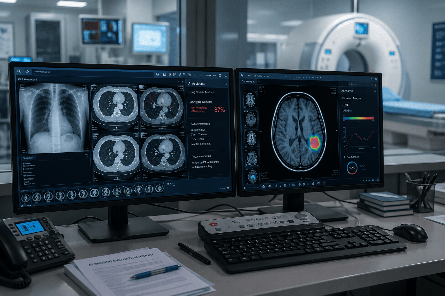
In 2025, the global AI medical imaging market was reportedly worth $37.9 billion. The forecast for 2026 clocks in at $52.9 billion, a 39.5% year-on-year jump1. The FDA is clearing more than 80 AI-enabled imaging devices every quarter. The industry narrative is clean and simple: AI is transforming radiology.
But if you're the person who actually has to decide what imaging equipment to buy next year — whether it's a CT scanner, an MRI upgrade, or a refurbished C-Arm — you don't need another market projection. You need to know whether these AI tools work in real hospitals, on real patients, under real constraints.
We went through the clinical evidence published in 2026. The answer is considerably more complicated than the press releases suggest.
The Numbers Nobody Agrees On: Why AI Imaging Market Forecasts Are All Over the Map
Start with two data points from the same market, same year.
The Business Research Company's February 2026 report pegs the 2025 global AI medical imaging market at $37.9 billion, projecting $52.9 billion for 20261. Fortune Business Insights estimates 2025 at just $18.8 billion2.
Same market. Same year. A difference of nearly 2×.
This isn't a rounding error — it's a fundamental disagreement about what counts as "AI medical imaging" and how to measure it. One firm may bundle AI analysis software, imaging hardware, cloud compute, and storage services into a single estimate. Another may count only pure software licensing revenue. Both reports are proprietary commercial research; neither discloses its methodology in full.
⚠️ Watch Out: The "AI imaging market will reach $XX billion" figures most often cited at industry conferences and in vendor pitch decks are almost always the upper-bound projections. When you hear a number, ask three questions: What's in the basket? Which firm produced the estimate, and from what report? Is it audited data or a model-based forecast?
The regulatory side offers a somewhat more grounded reference point. According to Innolitics' ongoing tracking of the FDA database, 86 AI/ML-enabled medical devices received clearance in Q2 2026 — 84 via the 510(k) pathway and 2 via De Novo classification3. That's a modest decline from 91 in Q2 2025 (-5.5%), and roughly in line with Q1 2026's 81 clearances — a steady-state, industrialized approval pipeline.
Here's the catch: FDA 510(k) clearance requires demonstrating "substantial equivalence" to an already-marketed device. It does not require proving clinical superiority over a baseline without AI. Those 86 clearances tell you the regulatory pipeline is functioning. They don't tell you whether any of these products, deployed in an actual hospital with actual patients, produce better outcomes than a radiologist reading images unassisted.
93,000 Patients, One Uncomfortable Answer: The LungIMPACT Trial
In March 2026, Nature Medicine published results from LungIMPACT — the largest randomized controlled trial of AI chest X-ray prioritization ever conducted: 93,326 X-rays, 86,945 patients, across 5 NHS trusts, from July 2023 through December 20244.
The design was straightforward: patients were randomized to either AI-assisted triage (Qure.ai's qXR v4.0 algorithm flagged suspected lung cancer cases as "urgent") or standard-of-care workflow. The primary endpoint was median time to lung cancer diagnosis.
The results deserve a careful read from anyone evaluating an AI imaging investment.
The AI did speed up one thing: report turnaround dropped from 47 hours to 34 hours (p<0.001) — a 13-hour improvement. But median time to diagnosis did not budge: 44 days in the AI arm versus 46 days in the control arm, p=0.844. That 13-hour gain upstream was entirely absorbed before it reached the patient. It didn't propagate.
Why? Because after the report came out, patients sat in the same queue waiting for a CT appointment. Median time to CT was identical across both arms: 53 days (p=0.31). Referral rates, time to treatment initiation, cancer stage at diagnosis — none of these differed significantly between groups. Across the entire trial, only 172 patients received a same-day CT. And that number was the same with or without AI4.
💡 Expert Insight: The trial's chief investigator, Nick Woznitza, put it plainly: the bottleneck isn't in image interpretation — it's downstream, in the referral pathway. The AI flagged the concerning cases. Nobody was there to act on the flag. AI is a sensor, not an actuator. A faster sensor connected to an unchanged actuator does not make a faster system.
There's a telling counterpoint. A separate study published in NEJM AI in January 2026 — the South West London trial — paired AI chest X-ray interpretation with a radiographer-led same-day CT pathway. When the AI flagged an abnormality, a radiographer booked the CT on the spot, before the patient left the department. CT time dropped from 6.0 days to 3.6 days5. Same AI concept, but with one added ingredient: workflow redesign that closed the loop between detection and action.
What this means for buyers. If you're evaluating an AI imaging tool, don't stop at the vendor's internal accuracy curves. Ask: do you have multi-center RCT data at anything approaching LungIMPACT's scale? Which step in the clinical workflow does your AI accelerate — and can that acceleration actually propagate to a patient outcome? If the answer stops at "faster report turnaround," you may be buying a metric that looks good in a spreadsheet, not a system that changes what happens to patients.
🔎 If your facility is evaluating AI software to augment diagnostic imaging, remember: the hardware that generates the images is still the hard bottleneck. Throughput and reliability of your CT, MRI, and X-ray equipment determine how fast any AI-assisted workflow can actually run. Search CT scanners on MedTrade and MRI systems by brand, model, and clinical application — send inquiries to multiple verified suppliers to compare configurations and lead times.
Lab Champion, Hospital Flunk: What Happens When AI Models Meet Patients They Weren't Trained On
This problem is subtler than "does AI work or not" — and more dangerous.
A 2025 systematic review aggregating hundreds of deep learning imaging studies reached a sobering conclusion: 81% of models showed significant accuracy drops when tested on external datasets — scanners they hadn't seen, hospitals they hadn't trained on, populations they hadn't encountered. Specificity fell by as much as 24 percentage points6. A model trained on Mayo Clinic data, deployed on a community hospital's aging CT scanner, can behave like a different piece of software altogether.
At the RSNA 2026 annual meeting, the ReclAIm framework was presented: of 18 deployed commercial AI models tested, 8 showed performance degradation, with the worst case dropping 40.6%7. FDA post-market surveillance confirms algorithm drift as a persistent threat — and here's the number that should concern anyone in procurement: of the 1,400+ AI-enabled imaging devices the FDA has cleared, fewer than 15% have published any real-world performance data7.
But the most vivid case study landed in July 2026.
Patwary and Toma, publishing in Applied Sciences, trained a ResNet-50 deep learning model for fracture classification. Under the standard setup — train and test on the same age group — the model achieved 82%–84% internal accuracy. That's a publishable result. It's also the kind of number a vendor might print in a product brochure8.
Then the researchers ran a test that ought to be routine before clinical deployment but almost never appears in academic papers: cross-age validation. Train on X-rays from younger patients (ages 20–35). Test on X-rays from older patients (ages 60–85).
Result: specificity collapsed to 0%8.
Zero percent specificity means the model flagged every single elderly patient's X-ray as a fracture — regardless of whether a fracture was present. Sensitivity was artificially inflated to 100%, but only because the model had learned a "flag everything" strategy, not because it had learned to diagnose. The researchers called it catastrophic failure, not gradual degradation8. Older bones have lower bone density, different trabecular patterns, narrower joint spaces — features underrepresented in the training set, which the model interpreted as pathology signals.
⚠️ Watch Out: When a vendor shows you an accuracy number, it's almost certainly from an internal test set — same hospital type, same demographic profile, same scanner generation as the training data. Ask for their external validation data: what populations? What age distribution? What equipment makes and models? How closely do those match your actual clinical environment? If they can't produce it, your procurement decision is a bet.
This pattern — stellar internal validation, unpredictable external performance — has direct implications for how you evaluate imaging hardware. If AI accuracy is sensitive to image acquisition parameters (and it is: one study found 69%–87% of chest X-ray AI performance variance is explained by technical acquisition differences8), then your scanner's image consistency matters more than ever. A lower-tier machine producing technically adequate but inconsistent images may undermine AI tools your department plans to adopt in the next five years.
When Large Language Models Look at Medical Images: GPT-4o's F1 Score Was 0.22
Since 2023, a popular narrative has held that general-purpose AI models are catching up to specialized medical imaging tools. GPT-4V can read an X-ray. Gemini can scan a pathology slide. The media coverage has been enthusiastic.
On July 3, 2026, Scientific Reports published Kukuljan et al.'s systematic evaluation of seven large vision-language models (LVLMs) across five cytomorphology benchmarks9. The narrative didn't survive contact with the data.
GPT-4o's zero-shot macro F1 score on peripheral blood cell classification was 0.22 ± 0.02 — close to random guessing. For comparison, a lightweight domain-specific hematology model (DinoBloom) substantially outperformed it9.
But the low score isn't even the most alarming finding. Two other results are worse.
First, the explanations were misleading. When experts reviewed the AI's classification rationales, 30% of GPT-4o's incorrect classifications were rated as "poor" or "misleading." The model wasn't analyzing the cell in front of it — it was regurgitating textbook descriptions of what that cell type "usually" looks like. When the image didn't match the textbook, the model delivered a correct fact incorrectly applied. It didn't lack knowledge. It lacked the ability to connect that knowledge to the specific image in front of it9.
Second, confidence was completely decoupled from accuracy. Fine-tuned GPT-4o's correlation between confidence and correctness was just 0.20. The zero-shot version: 0.03. Gemini-2.0: 0.05. Llama-3.2: 0.002 — functionally zero9. When these models tell you they're "95% certain" about a diagnosis, there is virtually no relationship between that number and whether they're right. In a clinical context, a tool that is highly confident and frequently wrong is more dangerous than one that knows when it's uncertain.
💡 Expert Insight: The researchers named this phenomenon the illusion of competence — the model produces fluent, plausible-sounding explanations on cases that resemble its training data, but fluency collapses when it encounters real, messy, out-of-distribution clinical inputs. The evidence as of mid-2026 is unambiguous: LVLMs should not be used for any diagnostic decision without human supervision. This isn't conservatism. It's what the data tells you.
A note of caution on interpretation: these findings are specific to cytomorphology tasks — cell-level classification. General-purpose LVLMs may perform differently on macroscopic radiological images. But the underlying pattern — high confidence paired with low accuracy on distribution shifts — echoes the generalization failures documented in dedicated imaging AI, and should give any clinical buyer pause.
Deep Dive: 15 Years, 1,879 Papers, and Only 21 That Bothered to Count the Money
Everything covered so far is about clinical performance. This section is about something arguably more important to a procurement decision: even when AI works clinically, can you demonstrate it's worth the check you're about to write?
Molwitz et al. published a systematic review in Radiology: Artificial Intelligence in 2026. They screened 1,879 AI imaging studies published between January 2010 and November 2024, spanning PubMed, Business Source Ultimate, and EconLit. The finding: only 21 — that's 1.12% — explicitly quantified any economic outcome10.
Not 21%. One-point-one-two percent.
An independent replication by Gregory et al., published in European Radiology in 2026, covered 2014–2025 and found 31 studies with any economic component at all, of which only 16 qualified as full economic evaluations (assessing both costs and outcomes)11.
Fifteen years. Thousands of papers. Almost nobody ran the numbers.
⚠️ Watch Out: This means that virtually any ROI claim you hear — "saves 30% of radiologist time," "20% efficiency gain," "18-month payback period" — has no independent, peer-reviewed economic evidence behind it. These figures come from vendor internal modeling, not from studies you can look up on PubMed. When the vendor says their tool pays for itself in 18 months, ask to see the independent study. If they can't produce one, it's because it doesn't exist.
Then there's the reimbursement side. In the United States, most AI imaging tools lack a dedicated CPT Category I code — cardiac CT is one of the few exceptions. This means they're bundled into the existing radiologist interpretation fee and cannot be billed separately to Medicare or commercial insurers7.
CMS data tells the story: between 2018 and 2023, Medicare received 83,392 claims for AI-related services. Approximately 47% were paid (total payouts around $8 million). Nearly 53% were denied (representing roughly $16.4 million in billed charges)7. Close to half of all AI service bills were rejected.
In Black Book's December 2025 flash poll of imaging leaders, 76% of respondents reported that AI "lacks clearly quantifiable ROI"7. When ROI can't be calculated in advance and the reimbursement pathway is unclear, procurement teams do exactly what you'd expect: they delay approvals, cap pilots at a single department, and shelve multi-site deployment plans.
This is why, despite hundreds of FDA clearances per year, most hospital AI imaging deployments remain stuck at the "research project in one department" stage. The clinical evidence is mixed. The economic evidence is nearly non-existent. And the billing pathway doesn't work.
What This Actually Means for Equipment Buyers
After five sections of clinical evidence, you might conclude that AI imaging is overhyped and not worth paying attention to.
The opposite is true. AI's impact on imaging medicine is real and significant — but it won't arrive through "replacing radiologists" or "making old scanners obsolete." It's working through a more subtle mechanism that directly affects equipment procurement decisions.
First, AI is increasing scan volume, not reducing it. When an AI flags more "suspicious findings," every flagged chest X-ray needs a confirmatory CT. Every incidental finding on that CT needs a follow-up scan. LungIMPACT's AI and control arms showed no difference in time to diagnosis — but the AI arm generated more downstream imaging demand. Scan volumes are rising. Equipment utilization is rising. Wear and tear is rising. Maintenance demand is rising. For imaging equipment buyers, this is structurally favorable: your hardware doesn't depreciate because AI arrived.
Second, AI is raising the bar for image quality — and that has procurement implications. If algorithm performance is sensitive to image acquisition parameters (and the evidence says it is), then entry-level equipment producing technically adequate but inconsistent images may not reliably support AI-assisted workflows. The question to ask isn't "can this scanner work with AI?" — it's "does this scanner produce images consistent enough for AI-assisted interpretation to be reliable?" That shifts the value proposition toward equipment with known, documented image consistency across patient populations.
Third, uncertainty itself is a decision framework. In 2026, with clinical evidence mixed and economic evidence nearly absent, the most rational equipment procurement strategy is: invest in hard-asset reliability — scanner throughput, maintenance response times, resale value — and treat AI as an optional incremental layer, not a decision foundation. Your capital budget should be allocated to things you can count on. Imaging hardware is something you can count on.
💡 Expert Insight: The refurbished and pre-owned imaging equipment market gains a new pricing anchor in the AI era. A 2020 GE CT, Siemens MRI, or Philips C-Arm produces image quality that fully supports current and near-future AI algorithm input requirements — at a fraction of new-equipment cost. In a transition period where AI ROI is unprovable and reimbursement pathways are unresolved, refurbished equipment offers a hedge: AI compatibility without the capital exposure of a new-system purchase.
Beyond the scanner itself, the accessories and components that keep imaging departments running — transducers, coils, tubes, detectors — follow the same logic. As scan volumes rise from AI-driven incidental findings, replacement cycles shorten. The procurement calculus for consumable imaging components becomes more favorable, not less.
One thing doesn't change no matter how AI evolves: you need actual imaging hardware to generate actual images before any AI — today's or tomorrow's — can do anything with them. From CT to MRI, from C-Arm to X-ray, the scanner is the non-negotiable first mile of AI-assisted diagnostic imaging.
On MedTrade, you can search for imaging equipment by brand, model, and clinical application — from CT scanners and MRI systems to X-ray equipment and components — compare supplier profiles, and send inquiries to multiple verified equipment providers. No AI can read an image that was never acquired. The hardware comes first.